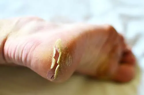
Cracked Heels
What causes cracked heels?
How should you treat cracked heels?
Cracked heels are very common and we see a lot of these in our podiatry clinic. You may feel embarrassed about seeking treatment for your cracked heels, especially if they’re not looking too great, but it’s important that you do. We can treat your heels, and help manage any pain and discomfort you’re experiencing, while they heal. We can also advise you on the best way to prevent cracked heels from occurring in the future.
Read on to learn more about cracked heels, what you can do to treat them at home, and how we can help if home treatment isn’t sufficient.
What are cracked heels?
Cracked heels are a very common foot problem. Most of the time, cracked heels are a merely unsightly to look at. However, if cracks (also called fissures) become large and deep, it can be painful to stand, walk or put any pressure on the heel. Left untreated, these cracks can also become infected.

signs and symptoms of cracked heels?
The first sign that you may develop a cracked heel is the formation of hard, dry callouses (patches of thickened skin) around the heel, which may be yellow or brown in colour.
As the skin becomes more dried out, these callouses can develop small cracks. Left untreated, these cracks will become larger and deeper as continued pressure is placed on the heel. Walking or standing or any other pressure on the heel will be painful. Over time, these cracks can become so deep that they may even bleed.
What causes cracked heels?
Cracked heels are caused by dry skin. When the skin around the edge of your heel becomes dry and thick, increased pressure on the pad under the heel causes the skin to crack. Cracked heels are more common during the winter months when humidity is low and the outdoor temperature is low. Other factors can also come into play including:
- Fungal skin infections
- Medical condtions eg. diabetes or psoriasis
- Medications
- Wearing open-heel shoes such as thongs or sandals.
- Barefoot walking
- Indoor heating which reduces indoor humidity and dries out skin
- Hot baths or showers that dry out skin

Who is at risk of developing cracked heels?
Anyone can develop cracked heels but there is usually a higher risk in people who have:

dry skin

eczema

juvenile plantar dermatosis

Diabetes

high bmi

palmoplantar keratoderma

psoriasis
Complications of cracked heels
Severe cases of cracked heels can lead to infection, including cellulitis (a common bacterial infection of the skin).
Cracked heels can also be serious for people who have neuropathic (nerve) damage. This can cause someone to lose feeling in their feet, thereby delaying treatment and increasing the risk of a serious complication. People with diabetes are at risk of complications such as diabetic foot ulcers.

Treatment for cracked heels
Fortunately, cracked heels can be treated successfully. However, the earlier you start treatment the better!
home treatment
Home treatments for cracked heels can successfully repair mild cases of cracked heels. These include:

Gently exfoliating your feet with a pumice stone or foot file to remove callouses.

Keeping heels covered by wearing breathable socks and avoiding open-heeled shoes.

Soaking feet in lukewarm water for up to 20 minutes to soften heels before exfoliating.

Moisturising the heels on a regular basis to hydrate skin. Look for heel balms containing urea as it helps remove dead skin. Be sure to moisturise after bathing or showering to help lock in moisture.
Podiatry treatment
In some cases, it may be necessary to see a podiatrist, particularly if home treatment is not making any difference, you are in a lot of pain, or the cracks in your heels are large. Podiatry treatment may involve:
- Debridement — cutting away the hard, thick skin (Note: only a qualified podiatrist should do this)
- Bandaging or strapping around the heel to reduce movement
- Insoles or padding to help redistribute the pressure on the heel
- In some cases, you may need prescription-strength ointment or antifungal cream. If we believe this is necessary, we can write the prescription for you
treat your feet to a medical pedicure
There’s no need to suffer from cracked heels in silence.
Our friendly, experienced podiatrists know exactly what to do to treat cracked heels, even in the most severe of cases.
Your Podiatrist will remove all dry skin and cracks around your heels. You will emerge from our clinic with hydrated crack free heels.
How to prevent cracked heels
As they say, prevention is better than cure and that’s certainly the case for cracked heels.
Fortunately, it’s relatively easy to prevent this annoying condition by following these steps:
- Check your heels each day for any signs of dryness or callouses
- Gently exfoliate any dry areas. You can even do this in the shower.
- Pat dry your feet after bathing
- Apply moisturiser on a regular basis to keep skin soft and supple
- Avoid wearing open-heeled shoes all the time.

Frequently asked questions
Removing the overlying dry skin and a good heel balm. In severe cases we recommend seeing a Podiatrist for foot care.
Yes, Podiatrists not only treat cracked heels but can also diagnose any underlying conditions.
A good heel balm should have urea as an ingredient. In some cases, creams which aren’t dense like a spray or mousse can absorb better into the skin.
At The Foot Hub we stock Restorate Foot Balm and PodoExpert Repair Foam
- Dry skin: Tips for managing. American Academy of Dermatology. https://www.aad.org/public/diseases/dry-sweaty-skin/dry-skin#overview. Accessed Feb. 18, 2019.
- Kermott CA, et al., eds. In Mayo Clinic Book of Home Remedies. 2nd ed. New York, N.Y.: Time Inc. Books; 2017.
- Litin SC, et al., eds. Skin, hair and nails. In: Family Health Book. 5th ed. Rochester, Minn.: Mayo Foundation for Medical Education and Research; 2018.
- Gibson LE (expert opinion). Mayo Clinic, Rochester, Minn. Feb. 18, 2019.
- Pawar M. The title of the paper: Treatment of painful and deep fissures of heel with topical timolol. J Am Acad Dermatol. 2020 May 29:S0190-9622(20)30975-0. doi: 10.1016/j.jaad.2020.05.100. Epub ahead of print. PMID: 32479981.
- IGNATOFF WB. Heel fissures and their management. J Natl Assoc Chirop. 1952 Feb;42(2):23-31. PMID: 14908525.
- Kelechi TJ, Lukacs KS. Patient with dystrophic toenails, calluses, and heel fissures. J Wound Ostomy Continence Nurs. 1997 Jul;24(4):237-42. doi: 10.1016/s1071-5754(97)90121-2. PMID: 9274281.
- Swensson O, Langbein L, McMillan JR, Stevens HP, Leigh IM, McLean WH, et al. Specialized keratin expression pattern in human ridged skin as an adaptation to high physical stress. Br J Dermatol. 1998;139(5):767–75. doi: 10.1046/j.1365-2133.1998.02499.x
- Rubin L. Hyperkeratosis in response to mechanical irritation. J Invest Dermatol. 1949;13(6):313–5. doi: 10.1038/jid.1949.47
- Kim SH, Kim S, Choi HI, Choi YJ, Lee YS, Sohn KC, et al. Callus formation is associated with hyperproliferation and incomplete differentiation of keratinocytes, and increased expression of adhesion molecules. Br J Dermatol. 2010;163(3):495–501. doi: 10.1111/j.1365-2133.2010.09842.x
- Thomas SE, Dykes PJ, Marks R. Plantar hyperkeratosis: a study of callosities and normal plantar skin. J Invest Dermatol. 1985;85(5):394–7. doi: 10.1111/1523-1747.ep12277052
- Farndon L, Barnes A, Littlewood K, Harle J, Beecroft C, Burnside J, Wheeler T, Morris S, Walters SJ. Clinical audit of core podiatry treatment in the NHS. J Foot Ankle Res. 2009;2:7. doi: 10.1186/1757-1146-2-7. ]
- Alavi A, Sanjari M, Haghdoost A, Sibbald RG. Common foot examination features of 247 Iranian patients with diabetes. Int Wound J. 2009;6(2):117–22. doi: 10.1111/j.1742-481X.2009.00583.x.
- Benvenuti F, Ferrucci L, Guralnik JM, Gangemi S, Baroni A. Foot pain and disability in older persons: an epidemiologic survey. J Am Geriatr Soc. 1995;43(5):479–84. doi: 10.1111/j.1532-5415.1995.tb06092.x.
- Dunn JE, Link CL, Felson DT, Crincoli MG, Keysor JJ, et al. Prevalence of foot and ankle conditions in a multiethnic community sample of older adults. Am J Epidemiol. 2004;159(5):491–8. doi: 10.1093/aje/kwh071
- Harvey I, Frankel S, Marks R, Shalom D, Morgan M. Foot morbidity and exposure to chiropody: population based study. BMJ. 1997;315(7115):1054–5. doi: 10.1136/bmj.315.7115.1054
- Spink MJ, Menz HB, Lord SR. Distribution and correlates of plantar hyperkeratotic lesions in older people. J Foot Ankle Res. 2009;2:8. doi: 10.1186/1757-1146-2-8.
- Pataky Z, Golay A, Faravel L, Da Silva J, Makoundou V, Peter-Riesch B, et al. The impact of callosities on the magnitude and duration of plantar pressure in patients with diabetes mellitus. A callus may cause 18,600 kilograms of excess plantar pressure per day. Diabetes Metab. 2002;28(5):356–61.
- Murray HJ, Young MJ, Hollis S, Boulton AJ. The association between callus formation, high pressures and neuropathy in diabetic foot ulceration. Diabet Med. 1996;13(11):979–82. doi: 10.1002/(SICI)1096-9136(199611)13:11<979::AID-DIA267>3.0.CO;2-A
- Reiber GE, Vileikyte L, Boyko EJ, del Aguila M, Smith DG, Lavery LA, et al. Causal pathways for incident lower-extremity ulcers in patients with diabetes from two settings. Diabetes Care. 1999;22(1):157–62. doi: 10.2337/diacare.22.1.157.
- Menz HB, Lord SR. Foot pain impairs balance and functional ability in community-dwelling older people. J Am Podiatr Med Assoc. 2001;91(5):222–9. doi: 10.7547/87507315-91-5-222
- Mickle KJ, Munro B, Lord SR, Menz HB, Steele JR. Foot pain, plantar pressures, and falls in older people: a prospective study. J Am Geriatr Soc. 2010;58(10):1936–194. doi: 10.1111/j.1532-5415.2010.03061.x
- Del Rosso JQ, Levin J. Clinical relevance of maintaining the structural and functional integrity of the stratum corneum: why is it important to you? J Drugs Dermatol. 2011;10(10 Suppl):s5–12. [
- Bikowski J. Hyperkeratosis of the heels: treatment with salicylic acid in a novel delivery system. Skinmed. 2004;3(6):350–1. doi: 10.1111/j.1540-9740.2004.04056.x.
- Goldstein JA, Gurge RM. Treatment of hyperkeratosis with Kerafoam emollient foam (30 % urea) to assess effectiveness and safety within a clinical setting: a case study report. J Drugs Dermatol. 2008;7(2):159–62.
- Akdemir O, Bilkay U, Tiftikcioglu YO, Ozek C, Yan H, Zhang F, et al. New alternative in treatment of callus. J Dermatol. 2011;38(2):146–50. doi: 10.1111/j.1346-8138.2010.00978.x.
- Potts RO. Stratum corneum hydration: experimental techniques and interpretation of results. J Soc Cosmet Chem. 1986;37:9–33
- Barba C, Méndez S, Roddick-Lanzilotta A, Kelly R, Parra JL, Coderch L. Cosmetic effectiveness of topically applied hydrolysed keratin peptides and lipids derived from wool. Skin Res Technol. 2008;14(2):243–8. doi: 10.1111/j.1600-0846.2007.00280.x
- Ciampi E, van Ginkel M, McDonald PJ, Pitts S, Bonnist EY, Singleton S, et al. Dynamic in vivo mapping of model moisturiser ingress into human skin by GARfield MRI. NMR Biomed. 2011;24(2):135–44. doi: 10.1002/nbm.1562
- Springett K, Merriman L. Assessment of the Skin and its Appendages. In: Merrimen MM, Tollafield RT, editors. Assessment of the Lower Limb. London: Churchill Livingstone; 1995. p. 207
- Pham HT, Exelbert L, Segal-Owens AC, Veves A. A prospective, randomized, controlled double-blind study of a moisturizer for xerosis of the feet in patients with diabetes. Ostomy Wound Manage. 2002;48(5):30–6.
- Baumgart E. Stiffness–an unknown world of mechanical science? Injury. 2000;31 Suppl 2:S-B14–23.
- Hashmi F, Wright C, Nester C, Lam S. The reliability of non-invasive biophysical outcome measures for evaluating normal and hyperkeratotic foot skin. J Foot Ankle Res. 2015;8:28. doi: 10.1186/s13047-015-0083-8
- Clarys P, Clijsen R, Taeymans J, Barel AO. Hydration measurements of the stratum corneum: comparison between the capacitance method (digital version of the Corneometer CM 825®) and the impedance method (Skicon-200EX®) Skin Res Technol. 2012;18(3):316–23. doi: 10.1111/j.1600-0846.2011.00573.x.
- Sans N, Faruch M, Chiavassa-Gandois H, de Ribes CL, Paul C, Railhac JJ. High-resolution magnetic resonance imaging in study of the skin: normal patterns. Eur J Radiol. 2011;80(2):e176–81. doi: 10.1016/j.ejrad.2010.06.002.
- Neto P, Ferreira M, Bahia F, Costa P. Improvement of the methods for skin mechanical properties evaluation through correlation between different techniques and factor analysis. Skin Res Technol. 2013;19(4):405–16.
- Feldman DL. Which dressing for split thickness skin graft donor sites? Ann Plast Surg 1991, 27(3): 288-91
- Pavicic T, Korting HC. Xerosis and callus formation as a key to the diabetic foot syndrome: dermatological view of the problem and its management. J Dtsch Dermatol Ges 2006, 4(11): 935-41
- Ahanchian N, Nester C, Howard D, Ren L. 3D modelling of the human heel pad. Salford Postgraduate Annual Research Conference 2012, 31-36. Available at: usir.salford. ac.uk/29427/1/2012_proceedings_v2.pdf
- Hashmi F, Nester C, Wright C, Newton V, Lam S.
- Characterising the biophysical properties of normal and hyperkeratotic foot skin. J Foot Ankle Res 2015, 12(8): 35
- Dockery GL. Mechanical Injuries. In: Cutaneous Disorders of the Lower Extremity 1997. WB Saunders, Pennsylvania, p242
- Iversen MM, Midthjell K, Tell GS, et al. The association between history of diabetic foot ulcer, perceived health and psychological distress: the Nord-Trøndelag Health Study. BMC Endocr Disord 2009, 9:18. Available at: http://bmcendocrdisord.biomedcentral.com/ articles/10.1186/1472-6823-9-18
- Bristow I. Emollients in the care of the diabetic foot. Diabetic Foot J 2013, 16(2): 63-6
- Cork MJ, Danby S. Skin barrier breakdown: a renaissance in emollient therapy. Br J Nurs 2009, 18(14): 872-7
- Penzer R. Emollients: selection and application. Podiatry Now 2005, 9: S1-S8
- Springett K, Deane M, Dancaster P. Treatment of corns, calluses and heel fissures with a hydrocolloid dressing. J Br Podiatr Med 2001, 52(7): 102-4
- National Institute of Health and Care Excellence. Type-2 Diabetes: Prevention and management of foot problems. Clinical guideline 10, January 2004. NICE, London. Available at: www.nice.org.uk/guidance/ cg10?unlid=93368616920162723333
- Young M, Townson M, Hicks G. A photographic scale to aid appropriate foot skin care for people with diabetes. Diabetic Foot J 2014, 7(2): 70
- Draelos ZD. Eczema Regimens. In: Draelos ZD (Ed), Cosmetic Dermatology: Products and Procedures, 2nd edn. John Wiley & Sons Ltd, Oxford, 2015
- Steel C, Longhurst B. An appraisal of IMUderm [Unpublished data]
- Beam JW. Tissue adhesives for simple traumatic lacerations. J Athl Train 2008, 43(2): 222–4
- Feldman DL. Which dressing for splitthickness skin graft donor sites? Ann Plast Surg 1991, 27(3): 288-91 17.
- Mertz PM, Davis SC, Cazzaniga AL, Drosou A, Eaglstein WH. Barrier and antibacterial properties of 2-octyl cyanoacrylate-derived wound treatment films. J Cutan Med Surg 2003, 7(1): 1-6
- Coulthard P, Esposito M, Worthington HV, van der Elst M, van Waes OJF, Darcey J. Tissue adhesives for closure of surgical incisions. Cochrane Database Syst Rev 2010, 12(5): CD004287
- Longhurst B, Allan E, Bristow IR. The use of cyanoacrylates in the management of dry heel fissures: a preliminary study. Podiatry Now 2010, 13(9): 11-15
- Vlahovic TC, Hinton EA, Chakravarthy D, Fleck CA. A review of cyanoacrylate liquid skin protectant and its efficacy on pedal fissures. J Am Col Certif Wound Spec 2011, 2(4): 79-85

A medical pedicure is your chance for that regular ‘check-up’, when a trained podiatrist will look after everything from dry skin to discoloured nails, callouses to corns, all in the hygienic setting of a medical clinic.

At The Foot Hub clinics we offer a cosmetic solution for damanged toenails.